

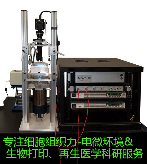
與成像分析系統(tǒng).jpg)

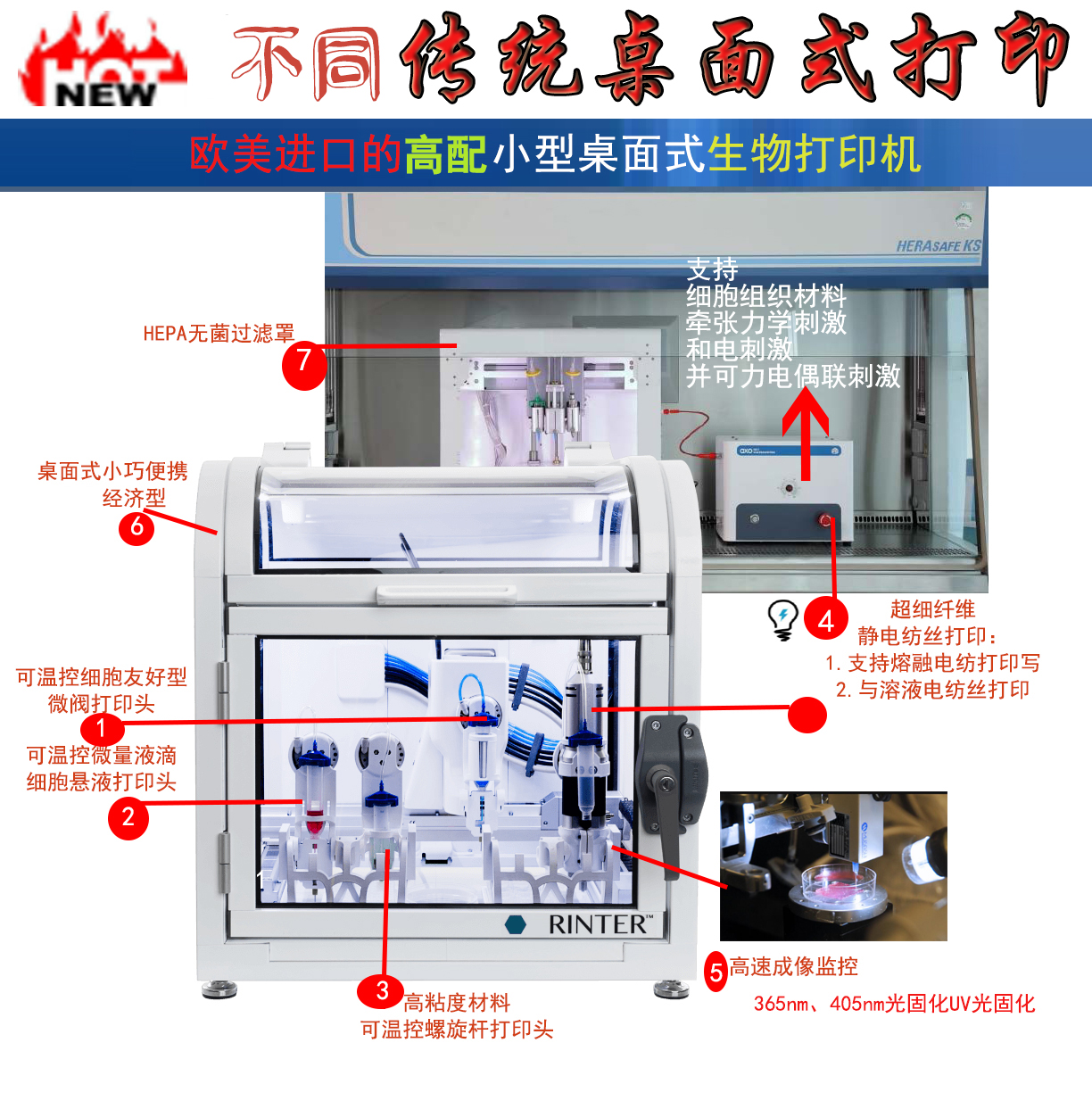
濟型溶體電紡絲生物3D打印機.jpg)
濟型高端生物打印機.jpg)

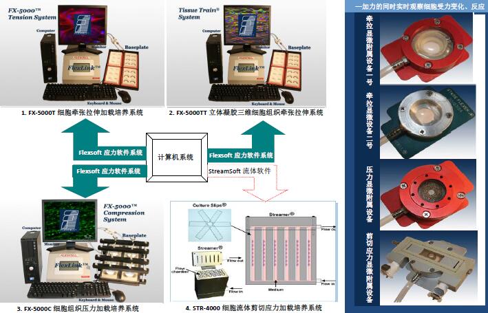
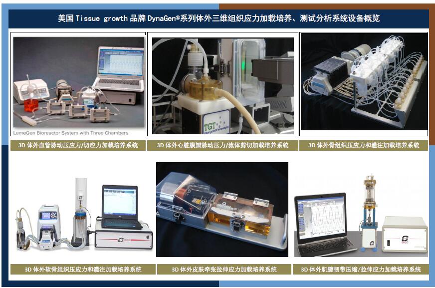
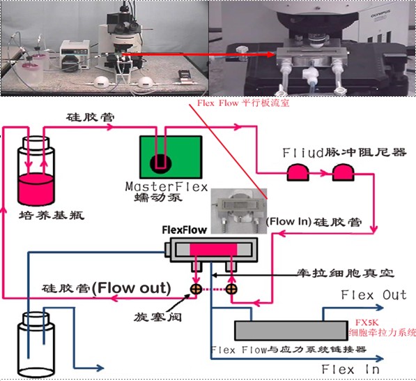
120-channel micro-electric array stretching stimulation and imaging recording system-force & electricity & imaging modular three-in-one
Enables researchers to reproducibly and reliably study the effects of physiological and pathological mechanical stretch on the electrophysiology of biological tissues. The system integrates:
(1)Cell stretch;
(2)Electrophysiological data acquisition system;
(3)Three functions of live cell imaging system。
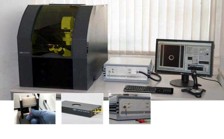
Optical Stretcher High Throughput Microfluidic Single Cell Mechanical Force Loading and Force Characterization Analyzer
In vitro vascular biomechanics experiment system
系統(tǒng).jpg)
-
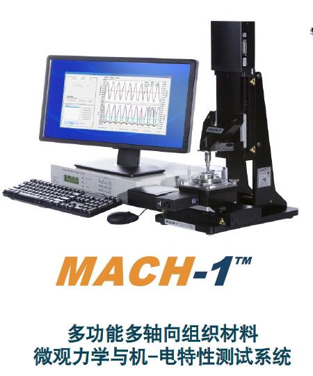
MACH-1 MECHANICAL TESTING SOLUTIONS FOR BIOMATERIALS AND TISSUES
Multifunctional biological tissue material compression, tension, shear, friction, torsion, indentation (stiffness and hardness), puncture, tear, push-out, expansion and contraction, bending, cantilever bending, blasting mechanical properties integrated test Analysis and cultivation equipment. It is limited to the measurement, characterization and evaluation of various mechanical properties of various materials.
● Can be placed in a standard incubator for incubation and supports multi-well plate testing
● Multiple biomechanical type testing and analysis functions
● Multiple materials: bone, cartilage, gel, skin, brain tissue, etc.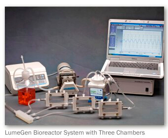
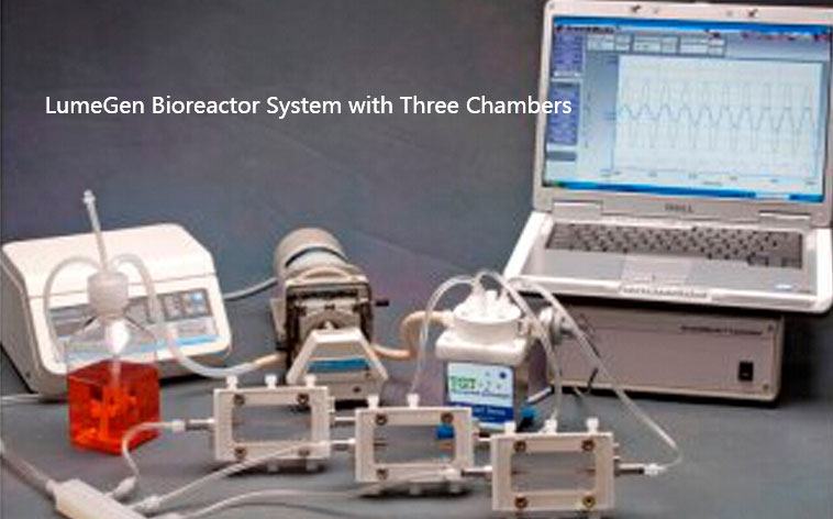
-TGT : Pulsatile Pressure & Flow Bioreactor System
This vascular in vitro stress culture system is a vascular in vitro culture device designed to help researchers perform proteomics of arteries cultured in vitro under strain, molecular signaling, effects of gene expression in vitro cultured vascular mesothelial smooth muscle cells, cellular and molecular mechanisms of aortic valve atresia and other genetic diseases in patients with aortic aneurysms, the effects of different stress conditions on in vitro cultured arterial mesothelial smooth muscle cell The in vitro stress culture system provides a variety of vascular connection tubes with different outside diameters to connect with vessels of different internal diameters, and the connection tubes are properly fixed by adding seals and fasteners. This solves the problem of coaxiality of the connection and fixation of the vessel, vessel connection tube and culture tank, and also solves the problem of sealing.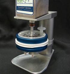
-TGT : Mechanical Compression Bioreactor Systems
The system is an ideal bioreactor for articular cartilage culture, mimicking the in vivo mechanobiological environment, transporting substances for cell growth and applying various mechanical stimuli to compensate for the inadequacy of existing in vitro construction techniques, providing a solution to the challenge of surgical repair of orthoarticular cartilage injuries.
-It is the ideal method for repairing damaged tissues by combining cytological and engineering principles.
-Provide compression, stretching, perfusion, and shock stimulation for disc-shaped samples such as cartilage
-Confocal real-time analysis, pressure perfusion mixed loading, multiple models for large samples to choose from
-3D culture and high-density inoculation
-The stability and controllability of material transmission, the exact control of stress magnitude, frequency, carbon dioxide and oxygen partial pressure, PH value, etc.,
-Simple and convenient operation, not easy to be polluted, long service life, etc.
-Growthworks? control system software: sinusoidal waveform capability, data acquisition and multi-motor operation control-to provide effective experimental models for orthopedic biomechanics and cartilage tissue engineering research.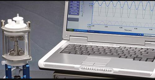
-TGT : Tension Bioreactor Systems
The system provides an oscillatory axial distraction and pressure stimulation culture environment for tendons and ligaments in vitro, and establishes a mechanical model of cell-degradable material complexes based on the biological and mechanical environment of tendon cells in vivo. Seeded cells were cultured and expanded and then inoculated on scaffold materials such as polyhydroxyacetic acid (PGA), serine protein, and polycaprolactone to form cell-material complexes and placed in a reactor for culture. Tendon tissues with good morphological and mechanical properties were constructed.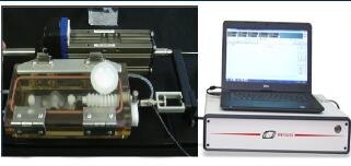
-TGT : Tension Bioreactor with Air/Media Interface
The system is capable of constructing tissue-engineered skin for different scaffold materials and cell complexes, simulating the growth environment and mechanical environment of skin according to the characteristics of skin growth and different scaffold materials and cell complexes, solving the problems of scaffold clamping and air-liquid interface in the construction of tissue-engineered skin through the bioreactor system, and providing a dynamic culture environment for a variety of skin cell complexes.
-TGT : Pulsatile Pressure & Flow Bioreactor System
Suitable for pulsation and shear stress stimulation of single valve structure. The standard structure has an inner diameter of 9 to 21 mm and a length of 50 to 80 mm. Modular design facilitates stent installation and cell Vaccination on stimulation system.
·Adapt to the vessels directly downstream of the two pulmonary valves and the vessels downstream of the oblique aortic valve.
·Light weight, compact structure, corrosion resistance, compatible with standard incubators
·Laser micrometer measurement, real-time measurement and monitoring, recording pipe diameter expansion rate, pressure
·Physiologically defined waveform control: static/forward rotation/cardiac/triangular/rectangular waveforms
·
-TGT OsteoGen: Perfusion Bioreactor Systems
The system applies oscillating axial stress and perfusion to cartilage constructs. It has single-chamber and multi-chamber configurations, and can be feedback controlled according to the properties of the material.
·3D cell and tissue culture to reveal the basic mechanisms by which cells perform their functions and guide the differentiation of stem cells
·Tissue characterization
·Development of biodegradable medical equipment
·Simulate the physiological conditions of disease models and pathological testing
·Optional sensors, non-contact micrometers, pressure sensors, etc., and/or modules
Customized instruments can add specific requirements to suit research applications.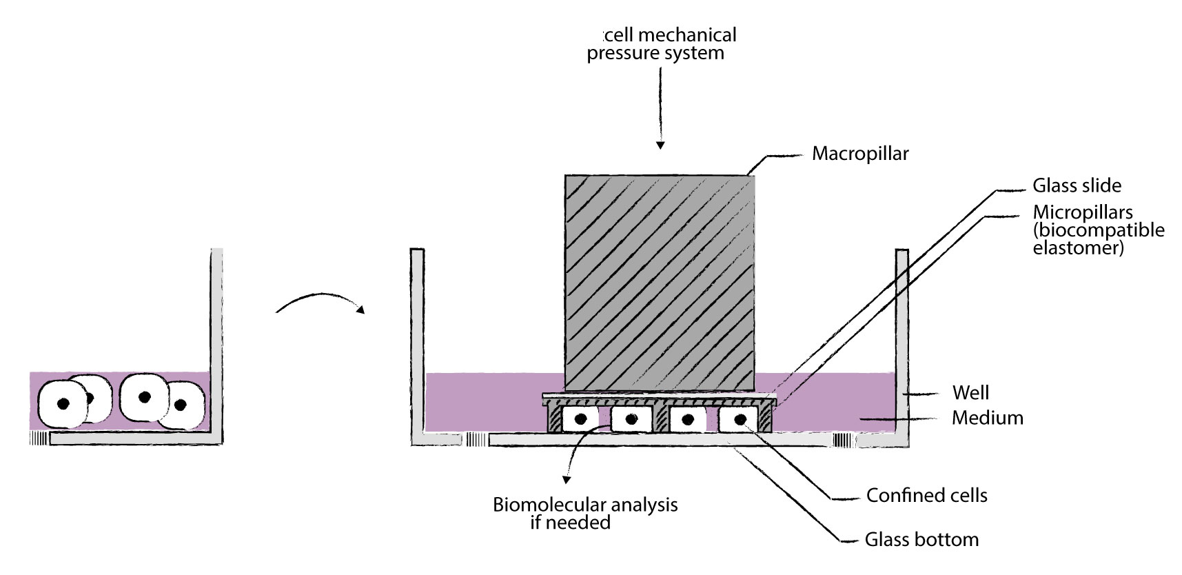
Single Cell Pressure Stimulation Culture System
Cells are uniformly confined/compressed between two parallel surfaces of two rectangular microcolumn arrays at submicron resolution. Different confinement heights (e.g. 1um - 300um),
● Allows long-term cell culture and cell proliferation while maintaining control of containment
● Compatible with high-resolution optical microscopy systems to handle a large enough number of cells for complete gene expression analysis, can be combined with biofunctionalized microstructured substrates and/or different substrates (geometry control) Use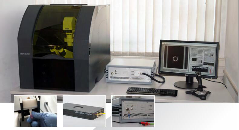
High-throughput single-cell mechanical force measurement analysis system
The system is an innovative laser optical tensioning platform technology for high throughput measurements of individual cell biomechanics. It is used worldwide to measure the deformability of individual cells in liquid at high throughput. The system is a module that can be mounted on any phase contrast microscope. Temperature stable and laser safe microscope system
1)Non-contact and label-free cellular measurements
2)High throughput - 250 cells/hr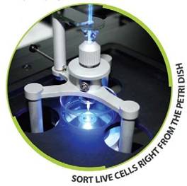
Automated cellular microtubule aspiration system
Model:Micropipette
1)Automatic computer control
2)Upgradeable configuration on existing microscopes
3)Can carry out cell grabbing and sorting, single cell separation and sorting, 1Cell/sec, can continuously sort 1000 cells at one time
4)Single cell adhesion, cell-to-cell adhesion measurement.
5)Single cells can be moved into each PCR tube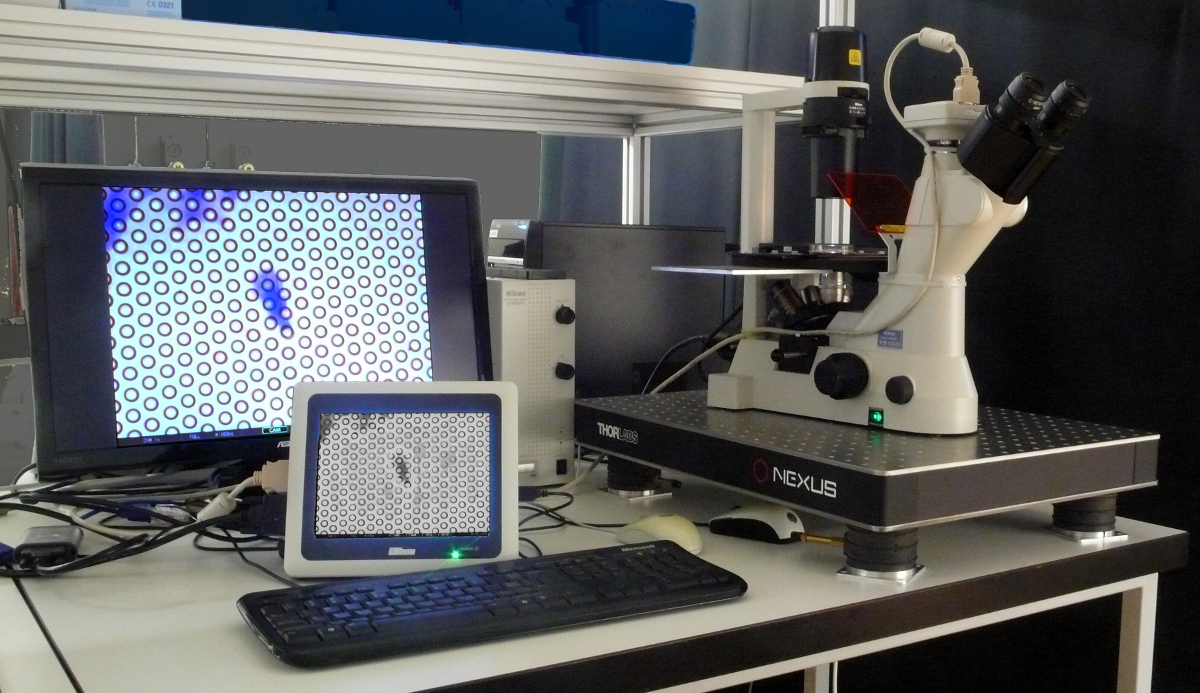
High-performance cell micropillar array traction microscope
Model:Cell traction microscope
The system reveals the close relationship between cell traction generated by the interaction of cells and extracellular matrix and cell proliferation, differentiation, apoptosis, embryonic development, tissue formation, wound healing, etc., and quantitatively studies cells at the single-cell level The temporal and spatial distribution of traction during migration (contraction) has important physiological and pathological significance
Cell microarray culture environment, imaging analysis of microcolumn deformation, quantitative analysis of cell traction
2)Upgradeable configuration on existing microscopes
3)Each array measures 3.2 x 3.2 mm and contains 10 x 18 observation points, each with 170 hexagonally arranged microcolumns
4)Micro-pillar elasticity range 1-3 nN (other requirements can be customized)
5)High-speed microarray imaging analysis software system, which can be used to extract cell mechanical parameters (force/microcolumn, microcolumn coordinates, microcolumn deformation, cell strain and stress distribution, etc.) from the cell pictures taken by the optical microscope. The analysis result can be saved as an Excel table for subsequent processing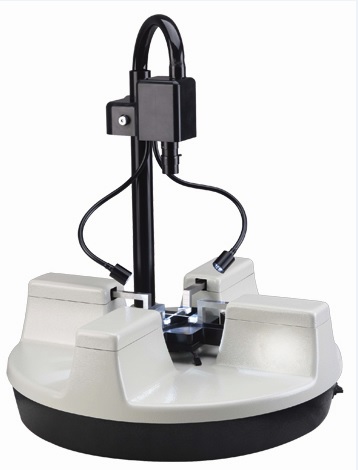
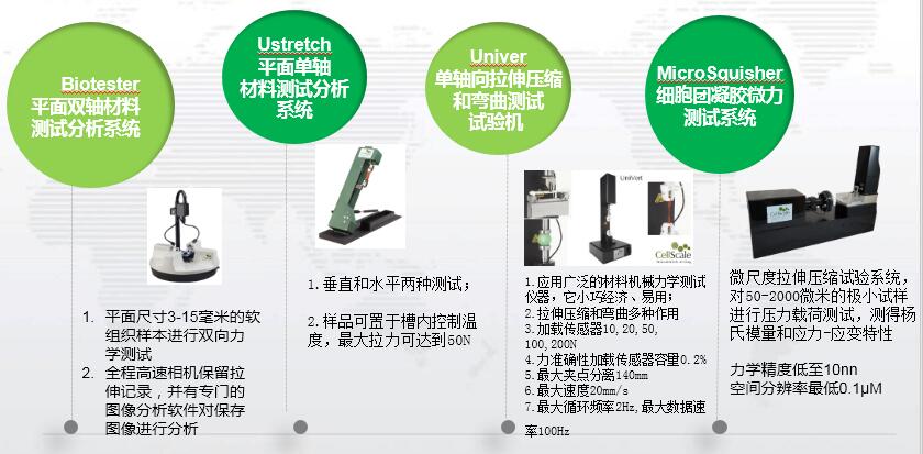
Cellscale Biotester Biaxial Tensile Test System
It is Cellscale's biaxial test system for soft tissue and biomaterials for anisotropy studies. It can capture dynamic images of the material and analyze the force and displacement process video at a later stage for analyzing the results and validation, and the resulting data can be easily exported to a standard spreadsheet for your experimental results at a glance! The range of applied materials includes: skin, ligaments, blood vessels, heart valves, sclera, cell membranes, scaffolds, etc. and their alternative bionic products. Data can be exported as Excel and imported into analysis software for analysis.
·1. Fast, accurate and repeatable sample loading; can perform uniaxial or biaxial tensile measurement of planar tissue;
·2. The test sample can be as small as 3X3 mm and as large as 15X15 mm; multi-module periodic, simple and relaxation tests can be performed;
·3. Real-time data mapping, comprehensive image tracking package, real-time feedback during testing and image analysis software; use the USB interface to quickly connect to the control computer with image and mark tracking software;
·4. Intuitive test design, integrated high-resolution CCD camera can provide simultaneous video tracking for dynamic imaging (up to 15 frames per second);
·5. Compact mechanical form factor;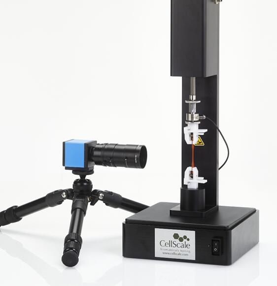
cellscale UniVert economical stretching, compression and bending universal testing machine
The UniVert universal testing machine is an ideal choice for various mechanical testing applications. Its small size and affordable price enable users to test at any time. The easy-to-use software and interchangeable component design make it easy to use it with a little training. The system can perform stretch, compression and bending tests with a force of 200N. Various fixtures and fixtures can be used to adapt to different specimens and test modes
Featured:
·The model is small and cost-effective.
·Fixtures and mechanical sensors are easy to replace and use to adapt to multiple purposes.
·Image strain measurement mode based on high-resolution CCD imaging (optional).
·Functional user interface software, realizing single, loop, relaxation and multi-modal testing, real-time feedback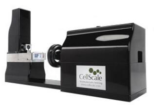
Cellscale MicroSquisher (Microtester) micro compression tester
The micro-mechanical performance test system can realize many functions that other instruments cannot. Smaller specimens, better force resolution, easier test setup and better visual effects. Applications include small tissue samples, hydrogel microspheres, cell spheres and engineered tissue growth substrates
Featured:
·Compression, tension, bending, indentation and shear test methods
·High-precision piezoelectric stepping device with 0.1μm resolution
·Force resolution as low as 10nN
·High resolution CCD imaging
·Integrated temperature control liquid pool
·Real-time feedback user interface software, easy to operate, can realize loop, relaxation and multi-modal tests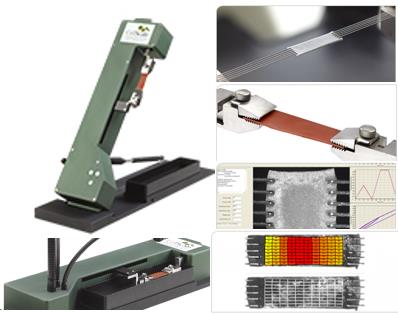
Cellscale Ustretch precision variable angle mechanical tester
The versatile precision single-axis benchtop mechanics tester is not limited to single-axis testing using a variety of sample fixtures. It is capable of performing vertical and horizontal testing in temperature-controlled media baths, as well as performing three-dimensional loading incubations of tissue samples. The system also features a variety of sample accessories, including screw-driven fixtures, spring-loaded fixtures and multi-point puncture fixtures.
Featured:
·High-performance actuator with built-in mechanical sensor
·High-resolution CCD imaging using image-based strain measurement tools
·Multiple accessory options, including the acquired BioRakes, for fast and reliable sample installation
·Integrated temperature control medium bath
·Full-featured user interface software for simple, loop, relaxation and multi-modal testing through real-time feedback
·The obtained BioRake accessory system can quickly and accurately install samples.
·The mechanical handle connection system helps to improve strength and testing.
·The integrated software can easily specify the test protocol, whether simple or complex. The test protocol can be saved and modified. Real-time force and displacement graphs help test verification.
·Optional imaging system and analysis software allow analysis of images to verify sample strain and strain uniformity.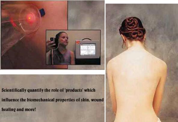
Srli BTC-2000 Skin flexibility and elasticity test and analysis system
The skin elasticity and other biomechanical properties of real-time quantitative test analyzer can be in vivo (In Vivo) or ex vivo (in vitro) non-invasive, real-time, quantitative analysis of skin, soft tissue, elastic materials, such as elasticity, relaxation, hardness (modulus), energy absorption and other biomechanical characteristics, from a biomechanical multi-angle quantitative measurement to characterize the ability of pharmaceutical formulations to change the biomechanical properties of the skin, as It is the basis for the design principles and efficacy evaluation of anti-aging and anti-wrinkle cosmetics.
-
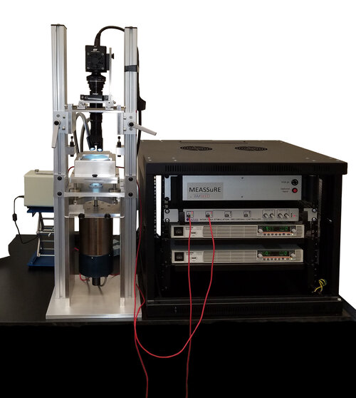
120-channel microelectric array stretching stimulation and imaging recording system
1)This cellular tissue microelectrical array stretch stimulation and imaging recording system allows researchers to reproducibly and reliably study the effects of physiological and pathological mechanical stretch on biological tissue electrophysiology. The system integrates:
(1)Cell stretching equipment;
(2)Electrophysiological data acquisition system;
(3)Three functions of live cell imaging system。
Each module can be used as a standing tool.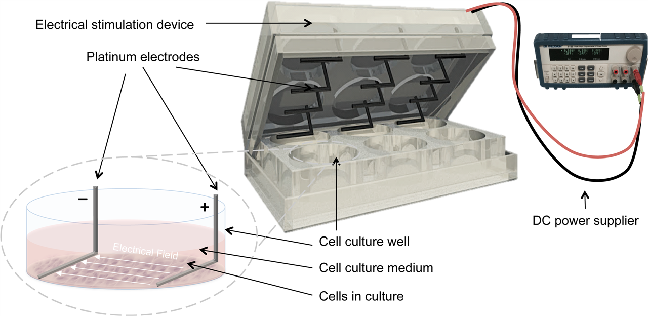
Cell Electrical Stimulation Culture SystemL-shaped platinum electrodes
Model:L-shaped platinum electrodes
The cell electrical stimulation culture system has been attracting much attention and has been widely used in promoting fracture healing, nerve fiber growth, infectious trauma, promoting plant growth, stem cell differentiation, cell electrotransfer, etc. It can simulate the in vivo state of cells for experiments. This system mainly includes electric stimulation controller, 6-well plate culture chamber and other parts, easy to install, easy to operate.
3.System Highlights
1)High throughput, multiple samples can be measured in parallel at the same time
This system uses multiple empty culture chambers to set up the same conditions for simultaneous parallel testing of multiple samples.
2)Allows real-time observation of experimental cell status
The cell culture chambers are transparent, allowing real-time observation of the state of the cells during the experiment.
3)The culture chambers can be placed in CO2 incubators to avoid contamination and provide an environment
The incubation chambers can be put into the CO2 incubator, and the electrical stimulation controller is connected outside, not only to avoid contamination contamination, but also to ensure temperature and humidity.
4)Different parameters can be set at the same time
This system uses multiple empty culture chambers that can be set up with different condition parameters to process samples with different parameters simultaneously on one culture substrate.
5)Multiple experimental methods can be set up and stored
This system software has a storage memory function, which can be set to store multiple experimental parameters, which can be quickly extracted during the experiment and can identify the appropriate parameters according to the optimization criteria, and the stored experimental parameters can also be cross-used in different multiple culture plates.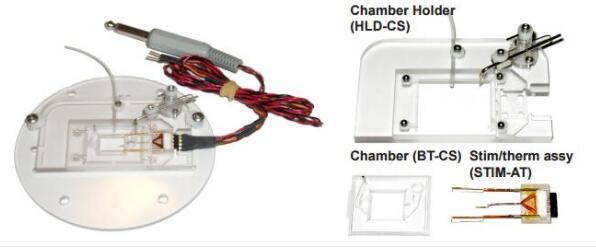
Cellular tissue electrical stimulation culture and real-time observation system
Model:Microcontrol
The cell stimulation system provides electrical stimulation of cells in culture to prevent their dedifferentiation, the culture chamber is reusable, the electrical stimulation is simultaneously perfused and can be observed and analyzed in real time by microscopy, and the special micro thermistor probe is fast temperature control to meet the cell culture requirements.
Features:
Low-cost reusable
In a typical installation with temperature control
Suitable for most popular microscope loading stages
Transparent bottom for real-time observation and analysis
Suitable for any stimulation
Perfusion can be performed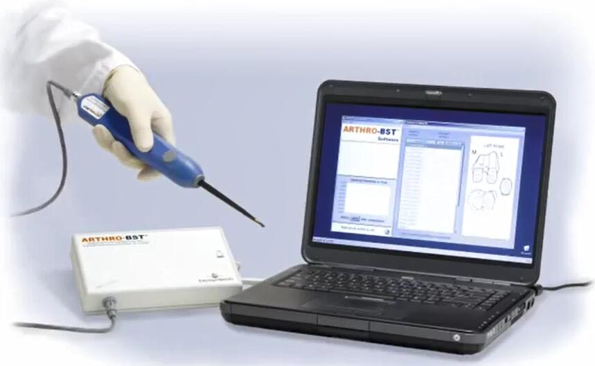
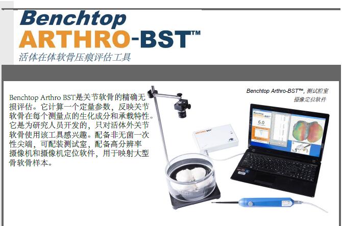
ARTHRO-BST Cartilage Isoflux Potential Test Analysis Evaluation System
Model:arthro-bst
The benchtop Arthro-BST measures compression-induced flow potentials of articular cartilage. These electrical signals are measured using an indenter during gentle compression of the cartilage surface. The special indenter design consists of a spherical surface covered with 37 microelectrical arrays. The device calculates a quantitative parameter of cartilage electromechanical activity (QP), which corresponds to the number of microelectricities in contact with the cartilage when the sum of its flow potentials reaches 100 mV. A high QP indicates weak electromechanical properties and vice versa. QP is reproducible and is independent of the applied force and indenter orientation.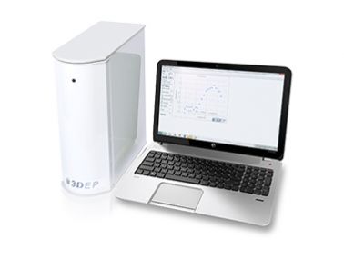
Dielectric electrophoresis cell characterization system
Featured:
1)Revolutionary New Technology:
The integration of microelectric microtiter chip, optical sensor detection system, automatic signal acquisition system and automated analysis software, etc., has commercialized the dielectrophoresis technology into the market for the first time; not only the operation threshold is lowered, but also the high stability and reproducibility, making dielectrophoresis cell analysis from theory to real and reliable application.
2)Chip Design:
The disposable chip design allows no chance of cross-contamination between experiments; the analysis of samples ranging from small viral particles to large cardiomyocytes is applicable, and the entire experimental sample does not need to be calibrated nor does it harm the sample.
Experiments are conducted with 20 holes at the same time with different frequencies, and results are available in just a few seconds, breaking away from the time-consuming and labor-intensive limitations of previous experiments.The 3D hole design can be used to quantify DEP-Force with a large number of samples and the change of lightness and darkness, making the stability and reproducibility of the experiment reach a commercial standard.
3)Automated analysis software:
The changes in lightness and darkness in the 20 pores are directly converted to DEP-Force, eliminating the need for complex physical formulas. With the parameters of environmental conductivity and electric field degree and size fixed, the software will automatically further calculate the cell membrane conductance, membrane capacitance, cytoplasm conductivity and cytoplasm permittivity), allowing more data to be obtained for the analysis of cells under dielectrophoresis.
Typical Applications:
1)Early monitoring of stem cell differentiation potential
2)Detection of early cell carcinogenesis
3)Rapid detection of bacterial drug resistance
4)Drug toxicity assessment
-
Chamber stand cell culture workstation
Designed for IVF cell culture and stem cell culture applications, this cell pH oxygen partial pressure CO2 partial pressure automation system manipulates the physicochemical environment (i.e. temperature, pH, osmolality, O2 and CO2 tension) for cell survival and development, providing a stable environment for culture in a precise gas and temperature environment. The in-line real-time pH monitoring system, with a good triple gas input, allows the connection of CO2, Nitrogen and/or premixed gases to the system, thus reducing costs and optimizing the incubator environment.
-Multiple compartmentalized chambers with each chamber parameter standing
-Quick recovery after opening the door to gas - less than two minutes after opening the door for 15 seconds
-Support three gas input!
-Fast anti-pollution!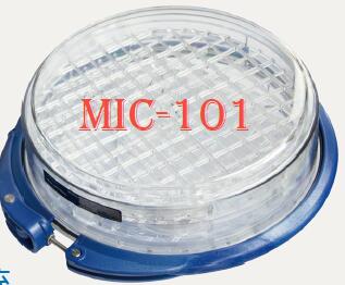
MIC-101 Economic Cellular Microbial Oxygen Strip Control Culture System
Featured:
Applications include in vitro fertilization, HIV isolation, establishment of hypoxic, hyperoxic and normoxic environmental conditions, organ culture and culture of large numbers of cell lines. Each unit is a stand-alone culture chamber that allows the establishment of the required culture chamber with good gas tightness and rapid gas exchange characteristics for fast and economical formation of a stable tissue culture environment (including gas concentration and humidity).
Microscope carrier cell gas control culture system
Featured:
1、Long-term imaging on a microscope stage (long-term live cell imaging and time-lapsed microscopy)
2、Suitable for any microscope carrier (Nikon, Olympus, Leica, mechanical carrier, confocal)
3、Mixable gas, oxygen deficiency control, precise control of oxygen and carbon dioxide concentration
4、Precise temperature control - cooling and heating control
5、Placement of culture containers such as slides and multi-well plates
6、Ultra-thin up to 150 micron
7、Flexible heater for any microscope objective lens
8、Instillable culture and fluid delivery
Cage type time difference microscope incubator
Featured:
The time-lapse microscopy is used to maintain the environmental conditions required for cell culture around the microscope workstation. The system platform is used to place multiple cell culture dishes or cell culture slides and to control the conditions of temperature, humidity, CO2 and air pressure to ensure proper cell growth, thus allowing for long time observation, long time culture and micro manipulation of biological samples. manipulation. The small size of the platform ensures that there is enough space for other equipment and that the cell proliferation rate in the cage microscope incubator is the same as in a conventional benchtop incubator.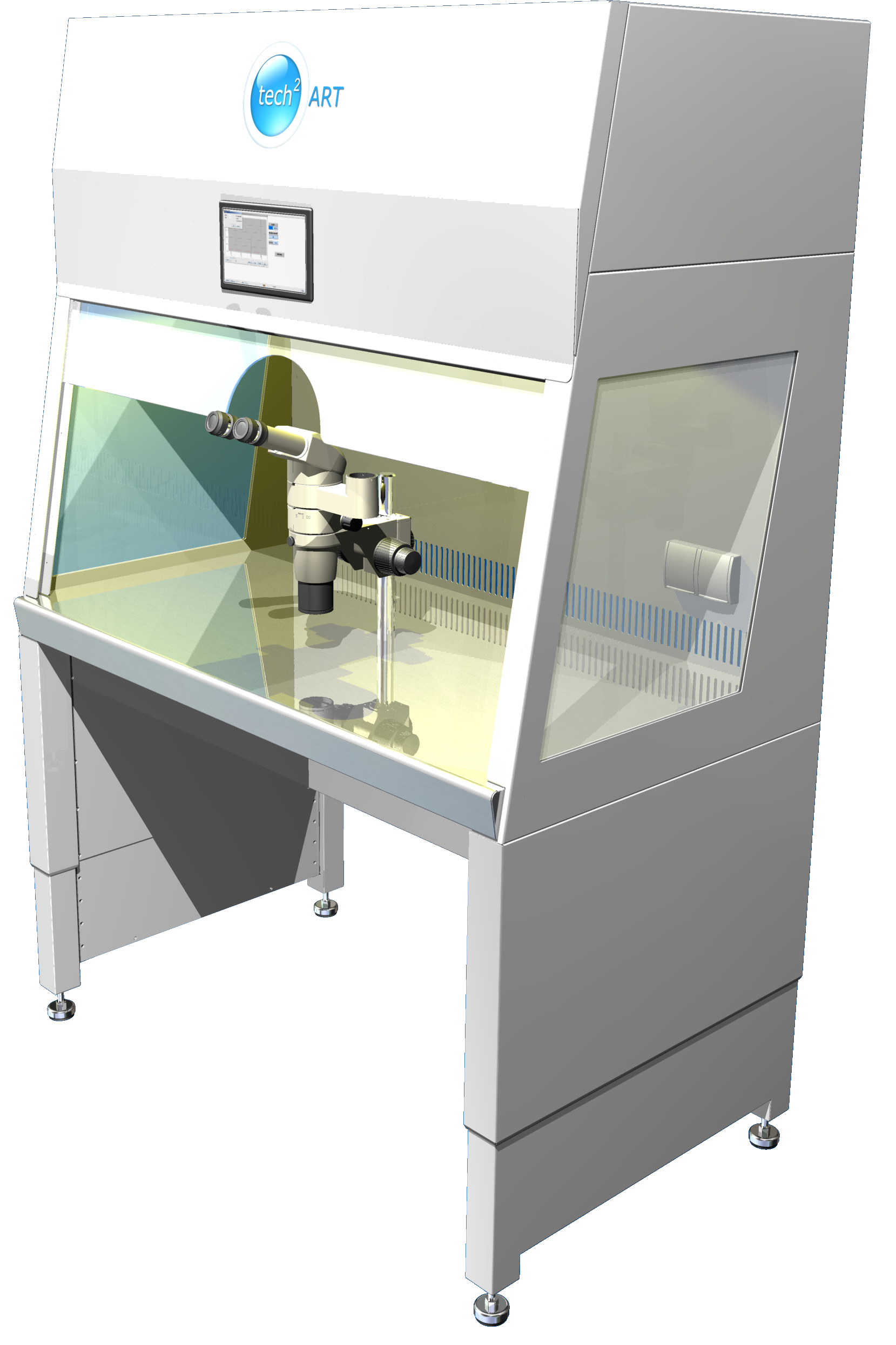
Time-lapse imaging, long-term incubation and real-time observation and analysis system for microscopy integrated with Class A II bio-safety cabinets
Featured:
1)Conditions your cell culture with closely monitored key environmental parameters to provide a high quality and consistent environment
2)Gas control, temperature control, PH control to provide environmental conditions for real-time observation and analysis of cell culture time differences
3)Microscope integration with Class A II bio-safety cabinets
4)Supports integration of any microscope, eliminating the need for duplicate microscope purchases
-
Automated high precision single cell grabbing and capture system
Model:SLBY_Cellsort
Featured:
The system is designed to automatically identify, pick and transfer single cells or single types of cells from suspensions with high precision, automatically and without affecting cell viability for reculture
1)Can upgrade an existing research-grade inverted microscope to a high-accuracy, high-throughput single-cell capture analysis system
2)Sorting speed: 1Cell/sec, 1000 cells can be sorted continuously at one time;
3)Single cells can be moved into each PCR tube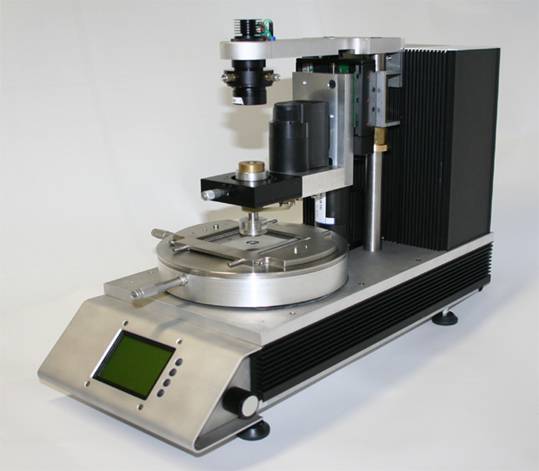
Single molecule manipulation analysis magnetic tweezer system
Featured:
The magnetic tweezer device is based on a dedicated inverted microscope, mounted on a motorized pan and rotate stage with a large set of single molecule manipulated magnetic tweezers.
The principle of the magnetic tweezer device is to observe and analyze the motion of the magnetizable beads by applying a force to them through a gradient-distributed magnetic field.
The magnetic tweezer device uses a pair of large rare-earth magnets (BdFeB) to enable the magnetization of the beads to reach saturation values.
Thanks to the application of a high-precision temperature control device, a special sample fixation device, a stepper motor to ensure the fast rotation of magnetic beads and a computer software system for flexible management, as well as a megapixel CCD camera, video acquisition and tracking measurements of up to 40 individual molecules can be achieved simultaneously, greatly improving the level of high-throughput analysis and statistics of single molecules.
Super-resolution force-sensitive 3D dual-optical tweezer system
Featured:
The super-resolution force-sensitive 3D dual optical tweezer system is a molecular biomechanical analysis system based on a dedicated inverted microscope, which combines optical tweezer technology and micro-visual image computation.
The system can be used as a stand-alone system in combination with a Zeiss Axiovert, AxioA1 or D1 microscope. The system is equipped with an infrared fiber laser with a power of 1 W or 5 W and can reach laser trapping forces in the range of 400 pN - 2 nN. The system's 3D-piezoelectric stage can achieve resolutions of 200 μn;m in the x- and y-axes and 20 μn;m in the z-axis direction. The Video-analysis system can achieve at least 2.5 nm lateral and axial resolution with an image capture rate of 200 frames/second and mutual imaging speed of 400 Hz in X, Y, and Z. It can analyze biomolecules in real time with 0.1 PN force resolution.
-
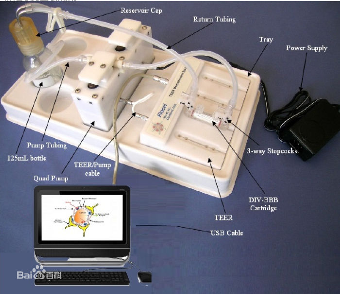
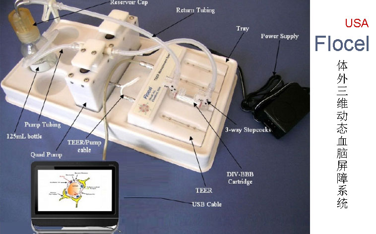
US in vitro dynamic 3D dynamic blood-brain barrier
Featured:
● More accurate pharmacokinetic and toxicological studies
● More accurate reflection of blood-brain barrier properties in vivo
● Mimicking important endothelial cell-astroglial cell interactions
● Electrical measurement of the integrity of the blood-brain barrier
● Ability to use real human cells
● Form tighter connections than existing static models
● Can significantly reduce drug development costs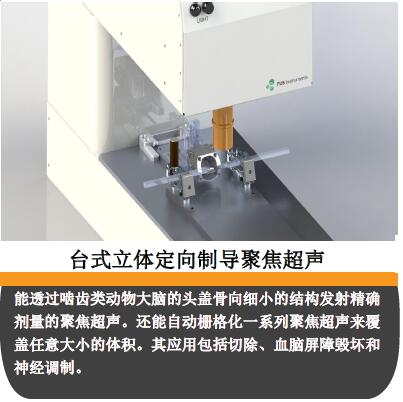
FUS Canada is the preeminent preclinical focused ultrasound technology company founded by researchers with more than 30 years of experience. Its two-photon microscopic imaging and focused ultrasound systems range from total solutions tailored for the introductory level to systems that allow users to go beyond the limits of focused ultrasound technology. It can help the world's scientists explore new drug delivery and development, as well as investigate new treatments for oncology, neuroscience and cardiovascular disease, allowing users to harness the research potential of focused ultrasound technology.
Canadian ultrasound fus tabletop focused ultrasound system
Featured:
1.Non-invasive, non-invasive: allows for longitudinal studies.
2.Precision: transmission of energy with a precision of millimeters or more.
3.One-stop system: image-guided treatment planning and preset dose parameters guarantee effective and reproducible results.
4.Accuracy: real-time energy monitoring and accurate acoustic calibration.
5.General: modulation of ultrasound parameters in the department.
6.Both acoustic energy and microscopic imaging analysis
Canadian ultrasound fus tabletop stereotactic guided focused ultrasound
Featured:
● This focused ultrasound system is capable of performing targeted brain exposure (opening the blood-brain barrier) without the need for concurrent imaging.
● The ability to emit a precise dose of focused ultrasound through the skull of the rodent brain to fine structures.
● It can also automatically rasterize a series of focused ultrasounds to cover volumes of arbitrary size. Applications include resection, blood-brain barrier disruption and neuromodulation.
● More accurate reflection of blood-brain barrier properties in vivo ● Mimicking important endothelial cell-astroglial cell interactions
● Electrical measurement of the integrity of the blood-brain barrier
● Ability to use real human cells
● Form tighter connections than existing static models
● Can significantly reduce drug development costs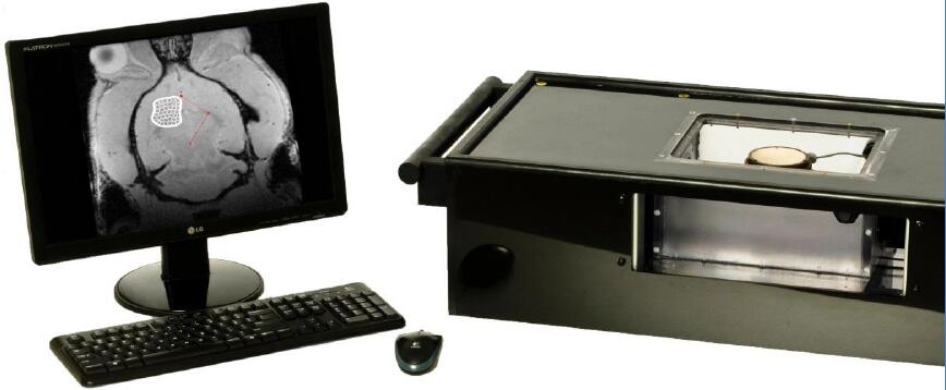
Canadian ultrasound fus-compatible MRI image-guided focused ultrasound system
Featured:
● Specifically designed for small animal models, it is a preclinical imaging system capable of non-invasive hyperthermia, resection and blood-brain barrier opening studies.
● The MRI-compatible image-guided focused ultrasound system consists of a computer-controlled, high-precision three-dimensional positioning system and a high-energy focused ultrasound transducer.
● The positioning system is capable of precisely delivering focused ultrasound energy to millimeter-sized areas to soft tissue.
● This system is specifically designed to study small to large animal models in order to investigate ultrasound-tissue interactions and to evaluate the safety of therapeutic approaches prior to use in humans.
● It can be matched to clinical MR and CT scanners for image-guided treatment planning and delivery. The system is completely non-magnetic and thus works with high field MRI and can also be matched to X-ray CT imaging.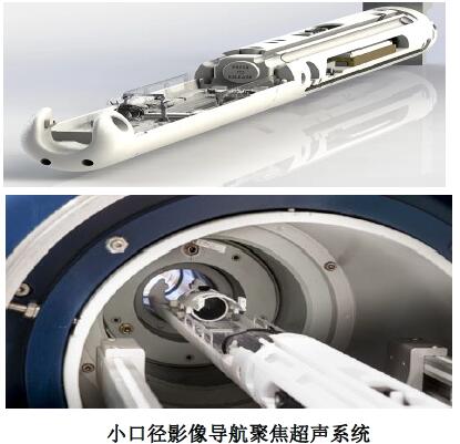
Canadian ultrasound fus small-bore image-guided focused ultrasound system
Featured:
● Specially designed for small animal models, it is a preclinical imaging system capable of performing non-invasive hyperthermia, resection and blood-brain barrier opening studies.
● Designed specifically for small animal models and suitable for blood-brain barrier disruption studies, it includes image navigation and positioning software, a computer-controlled high-precision two-axis positioning system and a calibrated focused ultrasound transducer.
●It is a preclinical imaging system capable of performing functions such as non-invasive hyperthermia, resection and blood-brain barrier opening.
Canadian ultrasound fus two-photon microimaging focused ultrasound brain exposure system
Featured:
● Industry microscope and focused ultrasound brain exposure system that allows simultaneous ultrasound and two-photon microscope visualization imaging, examination, and analysis.
● Designed specifically for small animal models and suitable for blood-brain barrier disruption studies, it includes image navigation and positioning software, a computer-controlled high-precision two-axis positioning system and a calibrated focused ultrasound transducer.
● It is a preclinical imaging system capable of performing functions such as non-invasive hyperthermia, resection and blood-brain barrier opening.

-
3D cell hollow rotating perfusion culture system a vascular tissue engineering reactor with culture lumen rotation, dual perfusion function inside and outside the vascular tissue, and intravascular nutrient fluid pulsation function, providing cell inoculation hollow and other scaffolds.
The system is a rotating perfusion bioreactor system with Silk Fibroin Tubular Scaffold (SF), Polycaprolactone Tubular Scaffold (PCL) and optical sensors for real-time monitoring of pH, oxygen and other parameters. and other culture parameters. The oxygen and nutrients in the vessel are mixed and delivered continuously to the microtubular channels of the scaffold, so that the cells adhering to the microtubular channels receive sufficient nutrients and are stimulated by a certain fluid shear stress to regulate the cell function. This system overcomes the disadvantages of static culture, improves the culture environment, increases the controllability of the culture process, and helps to promote the proliferation, differentiation and production of a large number of substrates by the cells adhering to the microtubules.
3D in vitro vascular biomechanics experimental system - in vitro 3D vascular pulse pressure fluid loading culture and analysis system
A vascular in vitro pulse compressive stress/shear stress incubation and monitoring analysis device, a bioreactor suitable for in vitro construction of tissue-engineered vessels, can accurately reproduce the in vivo environment of real blood vessels, and can provide a more scientific observation and evaluation of the long-term dynamic compliance and degradation performance of artificial blood vessels under simulated conditions.
1)Can be placed in an incubator for prolonged in vitro vascular pulsed compressive stress/fluid stimulation 3D culture
2)The pulse pressure and tangential stress can be adjusted separately to control the blood flow environment such as constant flow and pulsatile flow, and the pressure changes can be observed online to provide a more effective experimental means for the study of vascular biomechanics and vascular tissue engineering.
3)Provides realistic conditions that simulate the ambient temperature of the human body, the fluid environment outside the blood vessels, and the pulsating pressure from the continuous blood flow inside the blood vessels
4)Integrated CCD micrometer for measuring sample compliance, recording the rate of tube diameter expansion versus pressure in real time. 5)Transparent removable windows on both sides of the chamber allow optical monitoring with the instrument
Three-dimensional cartilage pressure and perfusion biomechanical system
The system is an ideal bioreactor for articular cartilage culture, mimicking the in vivo mechanobiological environment, transporting substances for cell growth and applying various mechanical stimuli to compensate for the inadequacy of existing in vitro construction techniques, providing a solution to the challenge of surgical repair of orthoarticular cartilage injuries.
-Physiological repair of damaged tissues by combining cytological and engineering principles is currently the ideal repair method
-Compression, stretching, perfusion, and shock stimulation for disc-shaped specimens such as cartilage
-Confocal real-time analysis, pressure perfusion mixed loading, large sample a variety of models to choose from
-Three-dimensional stereoculture and high-density inoculation
-Stability and controllability of material transport, exact control of stress magnitude, frequency, partial pressure of carbon dioxide and oxygen, pH value, etc.
-Simple and easy to operate, no pollution, long service life, etc.
-Growthworks? control system software: sinusoidal capability, data acquisition and multi-motor operation control - provides effective experimental models for orthopedic biomechanics and cartilage tissue engineering studies.
Large aspect ratio oscillatory compression/axial tension bioreactor system for three-dimensional tendon ligaments
The system provides an oscillatory axial distraction and pressure stimulation culture environment for tendons and ligaments in vitro, and establishes a mechanical model of cell-degradable material complexes based on the biological and mechanical environment of tendon cells in vivo. -Cell-material complexes were formed and cultured in a reactor using seeded cells cultured and expanded and inoculated on scaffold materials such as polyhydroxyacetic acid (PGA), filamentous protein, and polycaprolactone. -Construction of tendon tissue with better morphological and mechanical properties.
Three-dimensional skin-air-liquid interface tensor-stimulated culture bioreactor system
The system can be used to build tissue-engineered skin for different scaffold materials & cellular complexes. The system can simulate the growth environment and mechanical environment of skin according to the growth characteristics of skin and the characteristics of different scaffold materials & cellular complexes, and provide a dynamic culture environment for a variety of skin cellular complexes through the bioreactor system to solve the problems of scaffold clamping and air-liquid interface in the construction of tissue-engineered skin.
3D heart valve bioreactor (pulsatile flow pressure retraction stimulation)
Suitable for pulsatile and shear stress stimulation in single valve configurations. Standard construction sizes range from 9 to 21 mm inner diameter and 50 to 80 mm length. modular design allows for easy stent mounting and cell inoculation and stimulation systems.
·Accommodates two vessels directly downstream of the pulmonary valve and angled downstream of the aortic valve.
·Light weight, compact, corrosion resistant, standard incubator compatible
·Laser micrometer measurement, real-time measurement, monitoring and recording of pipe diameter expansion rate, pressure
·Physiologically defined waveform control: static/positive rotation/cardiac/trigonometric/rectangular waveforms
3D bone tissue culture bioreactor system
The system applies oscillatory axial stress and perfusion to cartilage constructs and is available in single and multi-chamber configurations with feedback control based on material properties.
·3D cell and tissue culture to reveal the underlying mechanisms by which cells exercise their functions and to guide the differentiation of stem cells
·Tissue characterization
·Development of biodegradable medical devices
·Simulation of physiological conditions in disease models and pathology testing
·Optional sensors, non-contact micrometers, pressure sensors, etc., and/or modules
Custom instruments can be added to suit specific needs for research applications
生產(chǎn)級中空纖維細胞培養(yǎng)系統(tǒng).jpg)
-
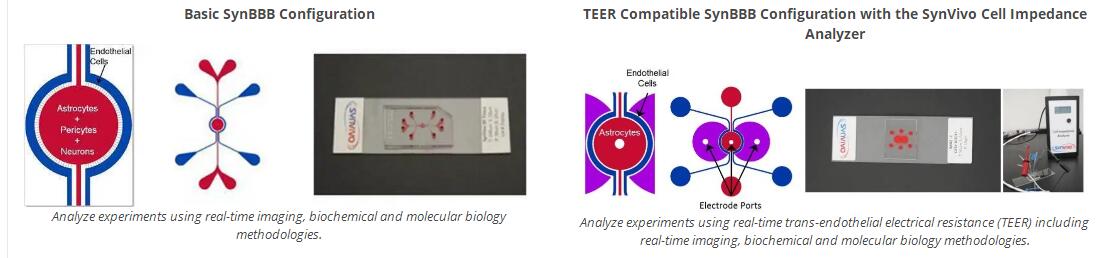
Synvivo Vascular Microenvironment and Pathology Cancer Model System
SynVivo a physiological, cell-based microfluidic platform that provides morphologically and physiologically very realistic in vivo microenvironments for real-time studies of cell behavior, drug delivery and drug discovery. SynVivo 3D tissue models reconstruct the complex in vivo microenvironment, including scale, morphology, hemodynamics and cellular interactions.
●SynBBB 3D blood-brain barrier model ●SynTox 3D toxicology model ●SynTumor 3D tumor model ●SynRAM 3D inflammation model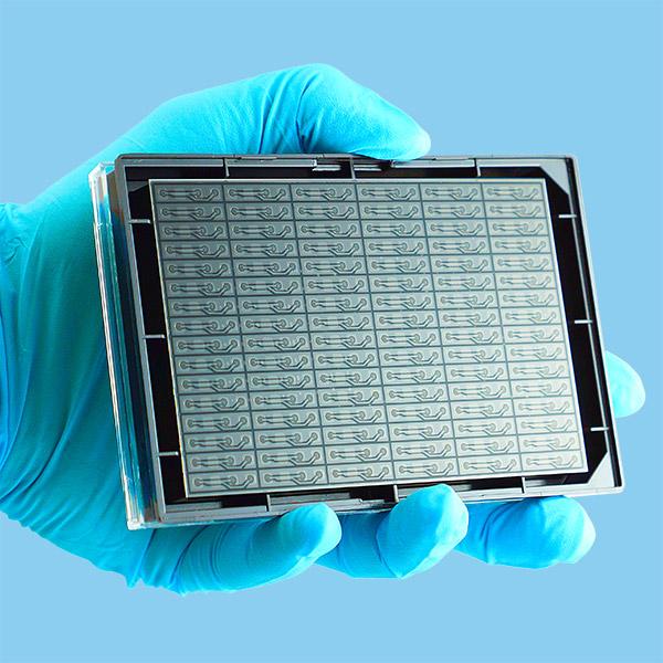
MIMETAS Organ-on-a-Chip (Organ Chip)
OrganoPlate is a microfluidics-based 3D cell culture plate that enables 96 single tissue models to be cultured on a single plate. Phaseguides enable the 3D redefinition of cells and their culture media, enabling cell-cell interactions and unprecedented imaging and quantitative manipulation. OrganoPlate? 2-lane, Item No. 9603-400-B; OrganoPlate? 3-lane, Item No. 4003-400-B
-
Economical PCB printing system
●Includes an inkjet print head that dispenses very fine conductive and insulating inks to create multi-layer rigid and flexible circuits on FR-4, Kapton or any substrate of your choice.
●Each printer has two heads for dispensing glue and solder paste, and another for picking up and placing components, allowing you to assemble the PCB on the desktop
●Multi-layer printing (up to 4 layers)
●Printing technology: thermal inkjet printing.
●Number of inkjet nozzles: 300
●Ink type (curing method): conductive (thermal), insulating (UV)
●Minimum trace width: 8 mils [200 microns]
●Minimum needle pitch: 16 million [400 microns]
●Minimum passage size diameter: 25 million [600 microns]
●Drill size: 15 million [400 microns]
●Maximum printable area: 4.6" x 6" [117 x 152 mm] [X / Y]
●Conductive ink thin layer resistance: 40 mOhms / square
●Supported formats: GERBER RS-274X,. jpg, .png, .tiff, .bmp
-
美國flexcell CellSoft微圖案化剛度可調(diào)節(jié)柔性基底膜細胞牽張和微圖案化剛度可調(diào)節(jié)玻片或多孔板
1)可在0.1-80kpa基底剛度可調(diào)節(jié)范圍內(nèi)牽張拉伸細胞
2)包被表面材料豐富:Amino, Collagen (Type I or IV), Elastin, ProNectin (R GD), Laminin (YIGSR).表面涂層豐富的包被材料, 您可以跟根據(jù)不同細胞組織可以靈活選擇不同包被材料表面節(jié)微圖案.jpg)
3基底剛度可調(diào)節(jié)微圖案培養(yǎng)玻片、皿、多孔板
法國微結(jié)構凝膠涂層玻片(兼容灌流)—可同時控制細胞形狀和剛度以重現(xiàn)體內(nèi)環(huán)境
●細胞在平坦或微結(jié)構化的軟3D環(huán)境中培養(yǎng),以模仿體內(nèi)條件。
●基于凝膠的基質(zhì)包含呈開放微通道(凹槽)或孔形式微圖案的、硬度可控制的(1-200kpa)微結(jié)構。
●可以在模擬體內(nèi)環(huán)境的微圖案特征和剛度的底物上培養(yǎng)細胞。
●預涂ECM基質(zhì)(例如纖連蛋白)。
●適用于任何細胞培養(yǎng)底物(蓋玻片,培養(yǎng)皿,多孔板)。
●凝膠的光學透明性使這些底物與高分辨率光學顯微鏡系統(tǒng)兼容
●隨時可用:直接接種細胞
●多種剛性模量:從非常軟(1 kPa)到非常硬(200 kPa)
●廣泛的3D設計:溝槽,方孔,圓孔等板,BioFLEX CULTURE PLATES.jpg)
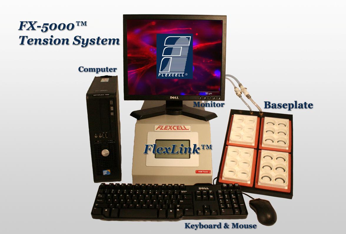
flexcell細胞拉伸應力加載培養(yǎng)板 CELL TENSION CULTURE PLATES--現(xiàn)貨
flexcell細胞拉伸應力加載培養(yǎng)板和FX-3000T/FX-4000T/FX-5000T/FX-6000T細胞拉伸應力加載系統(tǒng)配套使用,為細胞提供拉伸應力加載刺激培養(yǎng),包括
六孔 BioFLEX?雙向拉伸細胞培養(yǎng)板(6 well BioFLEX?CULTURE PLATES)
六孔 UniFlex細胞單軸拉伸培養(yǎng)板(6 well UniFlex Culture Plate)
24孔高通量BIOFLEX細胞雙軸拉伸培養(yǎng)板(24 well HT BIOFLEX CULTURE PLATES)
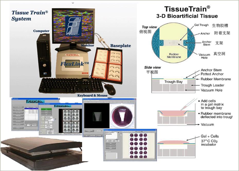
三維細胞或組織雙軸拉伸刺激培養(yǎng)板 (CIRCULAR FOAM TISSUE TRAIN CULTURE PLATES)
三維細胞或組織單軸線型拉伸刺激培養(yǎng)板 (Linear Tissue Train Culture Plate)
三維細胞或組織單軸梯型拉伸刺激培養(yǎng)板(Trapezoidal Tissue Train Culture Plate)
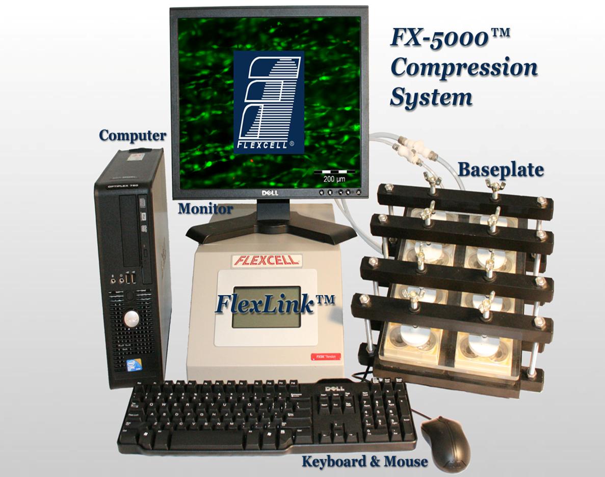
flexcell BIOPRESS細胞或組織壓力加載培養(yǎng)板 CELL COMPRESSION CULTURE PLATES--現(xiàn)貨
flexcell BIOPRESS細胞或組織壓力加載培養(yǎng)板和FX-5000C細胞或組織壓力加載系統(tǒng)配套使用,為細胞提供壓縮應力加載刺激培養(yǎng)。
六孔 BIOPRESS細胞或組織壓力加載培養(yǎng)板(BIOPRESS CELL COMPRESSION CULTURE PLATES)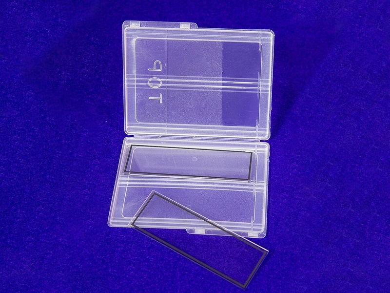
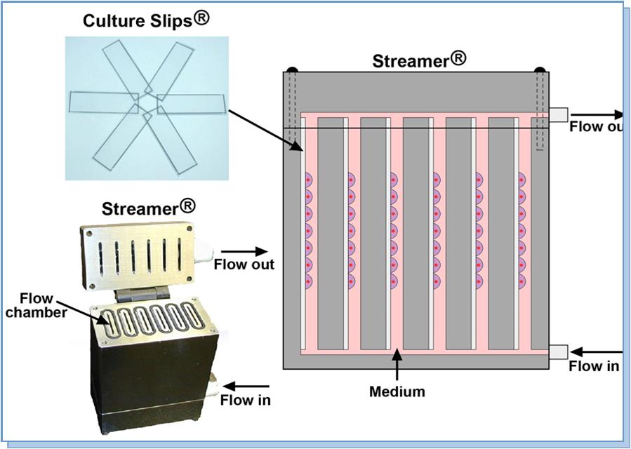
flexcell 細胞流體剪切應力加載培養(yǎng)玻片 Culture Slips Microscope Slides--現(xiàn)貨
flexcell 細胞流體剪切應力加載培養(yǎng)玻片和Str-4000細胞流體剪切應力加載系統(tǒng)配套使用,為細胞提供流體剪切應力加載刺激培養(yǎng)。
細胞培養(yǎng)載片包括顯微鏡載(物)片和蓋玻片兩種產(chǎn)品,表面經(jīng)過te殊處理,適合于細胞的貼壁與生長。
兩種規(guī)格:75 mm x 25 mm x 1.0 mm ,75 mm x 24 mm x 0.2 mm 。
75 mm x 25 mm x 1.0 mm 。
自身熒光低,光學性能佳。
不同包被的培養(yǎng)表面提高細胞的貼壁與生長。
五種不同包被的培養(yǎng)表面:Amino, Collagen (Type I or IV) Elastin, ProNectin (RGD), Laminin (YIGSR).
所以產(chǎn)品都是無菌du立包裝,僅供一次性使用。
訂貨信息
75mm x 25mm x 1.0mm 和 Streamer 或者 FlexFlow 配套使用
產(chǎn)品編號 英文名稱
CS-U Culture Slips — Untreated
CS-A Culture Slips — Amino
CS-C Culture Slips — Collagen Type I
CS-C(IV) Culture Slips — Collagen Type IV
CS-E Culture Slips — Elastin
CS-P Culture Slips — ProNectin
CS-L Culture Slips — Laminin
75mm x 24mm x 0.2mm 和 FlexFlow配套使用
產(chǎn)品編號 英文名稱
FFCS-U Culture Slips — Untreated
FFCS-A Culture Slips — Amino
FFCS-C Culture Slips — Collagen Type I
FFCS-C(IV) Culture Slips — Collagen Type IV
FFCS-E Culture Slips — Elastin
FFCS-P Culture Slips — ProNectin
FFCS-L Culture Slips — Laminin
-
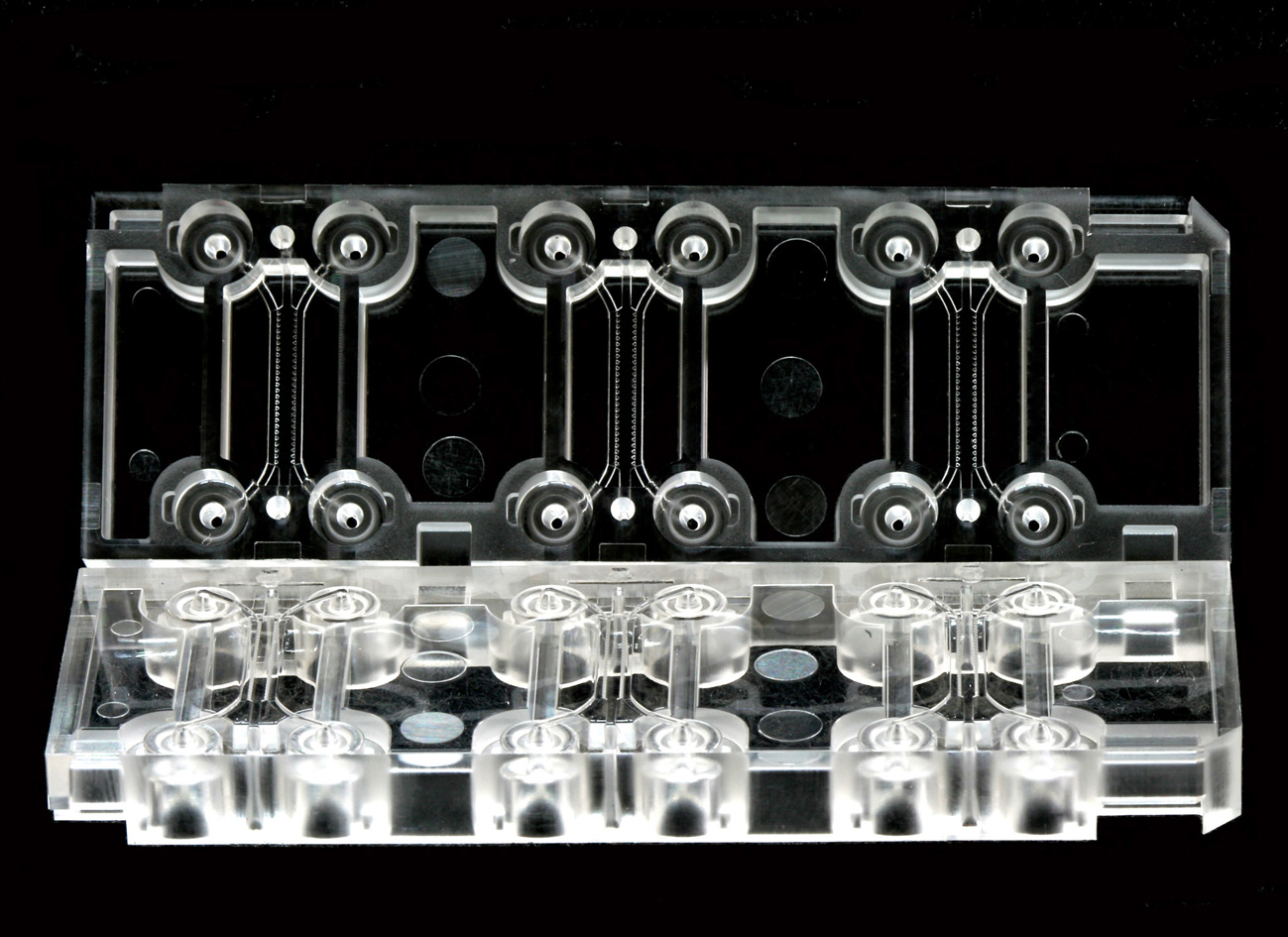 aimbiotech三維微流控培養(yǎng)芯片現(xiàn)貨
●1、Chipmate 3D細胞培養(yǎng)芯片采用三通道設計,中間為3D凝膠通道,兩側(cè)為培養(yǎng)基通道,通過負壓吸引快速交換培養(yǎng)基。
●2、芯片透氣性好,可有效進行氣體交換 3采用標準載玻片尺寸(75 mm × 25 mm),兼容相差顯微鏡、熒光顯微鏡和激光共聚焦顯微鏡
●4可實現(xiàn)不同類型細胞的共培養(yǎng)
簡介: 3D細胞培養(yǎng)芯片透氣性好,而且用戶可以通過選擇不同的水凝膠,在間隔的3D和2D空間進行不同類型細胞的培養(yǎng)。
同時可以通過對化學物濃度梯度和流體的調(diào)控很好地模擬符合用戶te定需求的微環(huán)境。
1、Chipmate 3D細胞培養(yǎng)芯片采用三通道設計,中間為3D凝膠通道,兩側(cè)為培養(yǎng)基通道,通過負壓吸引快速交換培養(yǎng)基。
2、芯片透氣性好,可有效進行氣體交換
3、采用標準載玻片尺寸(75 mm × 25 mm),兼容相差顯微鏡、熒光顯微鏡和激光共聚焦顯微鏡
4、可實現(xiàn)不同類型細胞的共培養(yǎng)
5、可在3D凝膠兩側(cè)產(chǎn)生化學梯度,也可控制3D凝膠中的間質(zhì)流
-
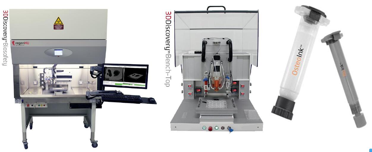
生物打印技術服務:
專注3D打印機、耗材及3D打印技術服務15年
為您量身定制解決方案,滿足廣泛的需求—設備銷售、租賃、科研技術委托服務
世聯(lián)博研專注生物力學和生物打印科研服務,10年經(jīng)驗支持。提供3D生物打印機銷售、租賃和3D生物打印科研實驗委托服務。世聯(lián)博研作為生物打印合作伙伴,為您提供解決方案——我們擁有專門的生物3D打印服務中心,配備多臺球的生物3D打印機,設備來自瑞士、美國、瑞典、芬蘭、法國、德國等。我們擁有一支由博士和碩士組成專業(yè)的技術團隊,公司在北京、廣州、上海、成都、合肥、西安、蘇州、哈爾濱、設立3D打印展示中心,為國廣大客戶提供現(xiàn)場觀摩和打印服務。我們專注您的業(yè)務,將您的想法帶入現(xiàn)實,我們幫您提供適合您行業(yè)的3D打印解決方案。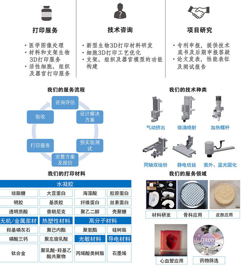
-
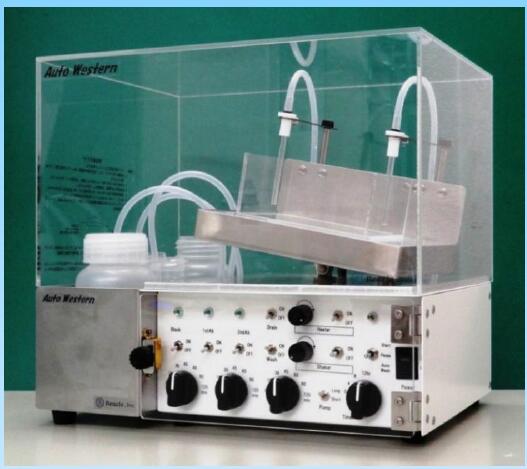
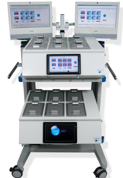
該自動免疫印跡試驗系統(tǒng)是用于進行膜的抗體反應以進行Western印跡的自動儀器。 抗體反應過程通常需要5個小時,使用Auto Western可以節(jié)省研究人員的勞力并減少錯誤。 此外,夜間使用該自動免疫印跡試驗系統(tǒng)可以地提高實驗效率。 使用Auto Western,膜抗體反應的手動過程可以自動進行。 它會按照以下順序自動換液:阻斷,清洗(3次),一抗,清洗(3次),二抗體和終清洗(3次)。 可以選擇反應時間。 該系統(tǒng)還利用了大多數(shù)研究人員都喜歡的搖動系統(tǒng)。 該系統(tǒng)旨在減少解決方案的殘留,因此靈敏度可與手動過程媲美。
特點:
●從阻斷到二抗體反應的自動化
●不需要te殊的反應室或試劑
●增靈敏度并減少反應時間(對于性病)
●高重復性
●操作簡便
●可用作蹺蹺板式搖床(用于迷你型)
●易于檢查操作過程
●省力省時
●可以使用通常的試劑和容器
●配備自動清洗系統(tǒng),易于維護
●定時器深夜無值守進行western blot
●抗體溶液由冷卻單元控制
●可以控制反應時間和溫度
●通過增加反應溫度和時間來節(jié)省抗體
●通過提高反應溫度來節(jié)省時間
●通過使用我們的增劑提高敏感性
BIO EXCELLENCE INTERNATIONAL Tech Co.,Ltd
